
MCL Injuries - Everything you need to know

Figure 1: Knee Pain - MCL Injuries - Physio Frenchs Forest, Physio Macquarie Park
What is the MCL
The knee has four ligamentous structures that act to stabilise the joint and limit excess movement. One of these ligaments is known as the Medial Collateral Ligament, or the MCL for short. The ligament runs over the inside of the knee, attaching the femur (thigh bone) and tibia (shin bone). The MCL also has some deep attachments into the meniscus of the knee, and for this reason meniscal and MCL injuries are often seen simultaneously. Due to its attachment points on the inside of the knee, the role of the MCL is to prevent the knee collapsing inwards (this is known as medial, or valgus collapse).
How do I injure my MCL?
Injuries to the Medial Collateral Ligament are the most common ligamentous injury seen in the knee joint, at around an estimated incidence of 40%. Unlike Anterior Cruciate Ligament injuries, damage to the MCL is usually sustained through direct contact to the knee. A blow to the outside of the knee, forcing it inwards causes the MCL to stretch, and potentially rupture completely. Other injuries associated with MCL injuries are meniscal tears and ACL sprains or ruptures.
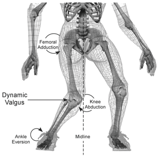
Figure 2: Knee Pain - MCL Injuries - Physio Frenchs Forest, Physio Macquarie Park
What are the symptoms of an MCL injury?
An injury to your Medial Collateral Ligament will usually result from a valgus type injury, as mentioned above. Pain, swelling and tenderness over the inside of the knee, where the thigh and shin bones meet is also common. Due to the swelling, your knee range of motion may be reduced, and you may struggle to weight bear on the affected leg. Finally, you may report to your physiotherapist that you felt a “pop” over the inside of the knee at the time of injury.
How is an MCL injury diagnosed?
The first step in diagnosing any knee injury is an assessment with a health professional, such as a physiotherapist or emergency physician. They will perform a number of physical tests to assess the integrity and stability of the ligament. Should your physiotherapist find it necessary, they can order an x-ray of the knee to assess for potential associated fractures. The gold standard of imaging for ligamentous injuries is an MRI, which can be requested to accurately assess the severity and location of your injury.
How are MCL injuries treated?
Treatment for Medial Collateral Ligament injuries is dependent on the grade of damage sustained. MCL injuries are graded from I to III, with grade I injuries involving a stretching of the ligamentous fibres. A partial tear of the MCL is considered a grade II injury, while a grade III injury describes a complete rupture of the ligament.
A grade I injury is treated with simple pain relief techniques, appropriate exercises prescribed by a physiotherapist, and a short recovery period (ranging from several days – two weeks).
Grade II and III injuries are treated slightly more conservatively, requiring bracing in specific positions to ensure correct healing, and to protect the ligament throughout this process. Grade II tears of the MCL are expected to heal over a 2-4 week period, while grade III ruptures require at least 6-8 weeks before returning to specific activities.
Surgery is rarely indicated for MCL injuries, although may be required if the ligament does not respond to conservative management. Tears or ruptures further towards the shin bone are less likely to heal completely than injuries to the upper portion of the ligament. A graft is taken from either the hamstrings, Tibialis Anterior muscle, or Achilles’ tendon when performing a Medial Collateral Ligament reconstruction.
See our expert Physiotherapy team at Frenchs Forest and Macquarie Park!